Parkinson's Drug Disrupts Gut Microbiome Due to Iron Deficiency

A new study, conducted within the framework of the FWF-funded Cluster of Excellence “Microbiomes drive Planetary Health”, and published in Nature Microbiology reveals how the Parkinson's drug entacapone disrupts the gut microbiome by inducing iron deficiency, favoring pathogenic microbes like E. coli.

The research, led by Michael Wagner from DOME in collaboration with CeMESS members and international partners from the UK, Denmark, and the US, highlights the often-overlooked effects of human-targeted drugs on microbial communities. Using a novel experimental approach, the team discovered that entacapone triggers iron starvation in the gut, promoting the growth of bacteria adapted to low-iron conditions. This groundbreaking study offers insights into how certain drugs can unintentionally alter the gut microbiome and suggests strategies to mitigate these effects.

NanoSIMS was used in the study to analyze the iron content in gut microbiota cells after exposure to entacapone. The method involved preparing faecal and bacterial cell samples, which were fixed, washed, and filtered onto gold-coated polycarbonate filters. These were then analyzed using a NanoSIMS 50L ion microprobe. The technique enabled the detection of secondary ion signals for elements such as carbon (12C+), sodium (23Na+), phosphorus (31P+), calcium (40Ca+), and iron (56Fe+). The study used NanoSIMS imaging to generate high-resolution elemental distribution maps, revealing that entacapone exposure increased iron accumulation in gut bacteria, particularly in Phocaeicola dorei. The relative iron content was quantified by normalizing the 56Fe+ signal to 12C+, allowing single-cell level comparisons.

Links

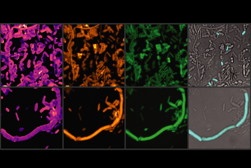
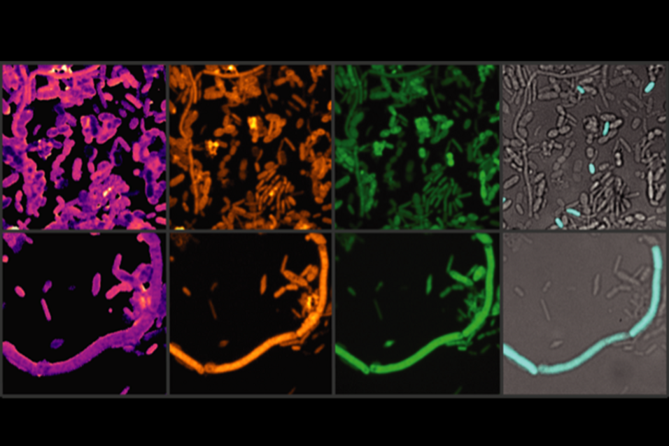
Chemical imaging of active gut microbes. After brief incubation with heavy water, culture medium and a drug, various chemical bonds (here C-D and C-H) in the stool sample are shown in yellow and green, their ratio in yellow-purple (left). Selected microbes are detected in the same image section with fluorescence-labelled oligonucleotide probes in cyan. The activity of the detected microbes can be determined based on the amount of C-D bonds.

Photo credit: Xiaowei Ge (Boston University)

 NanoSIMS News

20.11.2024
 

A new study, conducted within the framework of the FWF-funded Cluster of Excellence “Microbiomes drive Planetary Health”, and published in Nature...

05.11.2024
 

A new research project led by a team of four scientists, including CeMESS Senior Scientist Katharina Kitzinger, alongside researchers from Denmark,...

16.08.2024
 

This unexpected finding revealed a fourth growth substrate for ammonia oxidizers and offers new insights into niche partitioning of nitrifiers,...

19.04.2024
 

Desert soil microorganisms endure long droughts in a dormant state and rely on rain events for their reactivation. An international team around Dagmar...

13.03.2023
 

A research consortium led by Michael Wagner, deputy head of CeMESS, was selected in the competitive excellence program of the Austrian Science Fund...

30.07.2021
 

Three Divisions of CMESS now have a new home: Together with CUBE and TER – two of its sister divisions – DOME has moved to the new University of...