Nanoscale Secondary Ion Mass Spectometry Lab
at Large-Instrument Facility for Environmental and Isotope Mass Spectrometry, University of Vienna
Analytical nanometer-scale secondary ion mass spectrometry (NanoSIMS) imaging is perfectly suited to measure and visualize the distribution of virtually any elements and their stable isotopes in sample of various origin (from biology to cosmochemistry to material science). The CAMECA NanoSIMS 50L, which is available since 2010 at the Large-Instrument Facility for Environmental and Isotope Mass Spectrometry, offers a spatial resolution for elemental/isotope mapping down to 35 nm and thus even allows highly sensitive analyses at the sub-cellular level.
Services we provide
Beyond performing measurements, our facility offers extensive support to all users with respect to study design, sample preparation and pre-characterization, data evaluation and data interpretation, which are key-steps in an efficient NanoSIMS analysis. Applications for NanoSIMS projects within the fields of biology, chemistry, medicine, materials science and earth sciences can be submitted at any time. You can learn more about our cost model here.
Key research fields
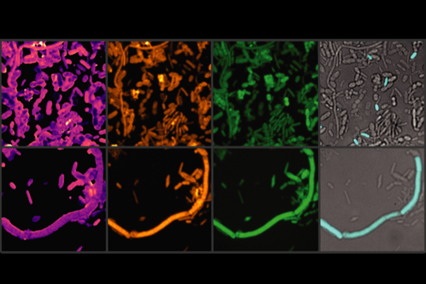
In microbial ecology we combine NanoSIMS with stable isotope probing (e.g. 2H/1H, 13C/12C, 15N/14N), high-throughput elemental analysis - isotope ratio mass spectrometry (EA-IRMS), Raman micro-spectroscopy and single cell identification techniques such as fluorescence in situ hybridization (FISH) for obtaining yet inaccessible information about the phylogenetic identity and functional role of microorganisms in their environment.
In collaboration with colleagues from the Department of Inorganic Chemistry NanoSIMS has been exploited for studying the sub-cellular distribution of isotopically-labeled metal-based anticancer drugs within two- and three-dimensionally cultured malignant cells as well as within normal and tumor murine tissues. In this context, combining NanoSIMS with fluorescence microscopy and quantitative laser ablation inductively coupled mass spectrometry (LA-ICP-MS) for millimeter-scale chemical mapping and transmission electron microscopy (TEM) for single cell ultrastructure characterization has proven successful.


